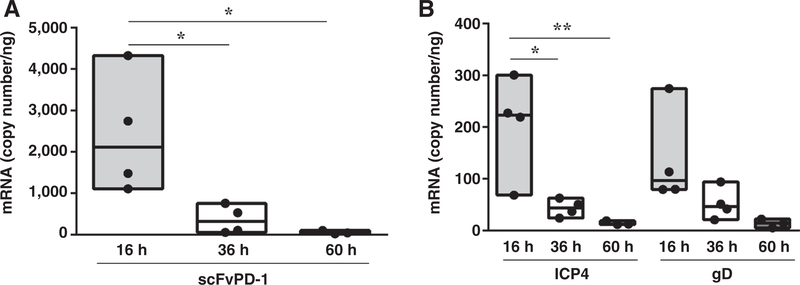
Figure 5.
In vivo viral gene and scFvPD-1 expression after GBM treatment with NG34scFvPD-1. C57Bl/6 mice were intracranially injected with GL261N4 cells (105 cells). After 7 days, NG34scFvPD-1 was intracranially administered at a dose of 1.5 × 10⁶ pfu/mouse. Sixteen, 36, and 60 hours after virus inoculation, mice were sacrificed, their brains harvested, and RNA isolated. scFvPD-1 (A) and immediate early gene (ICP4) and late gene (gD) mRNA copy number were assessed by real-time PCR (B). One-way ANOVA (Tukey multiple comparisons test) was used for the analyses (*, P < 0.05; **, P < 0.01).

