Figure 4.
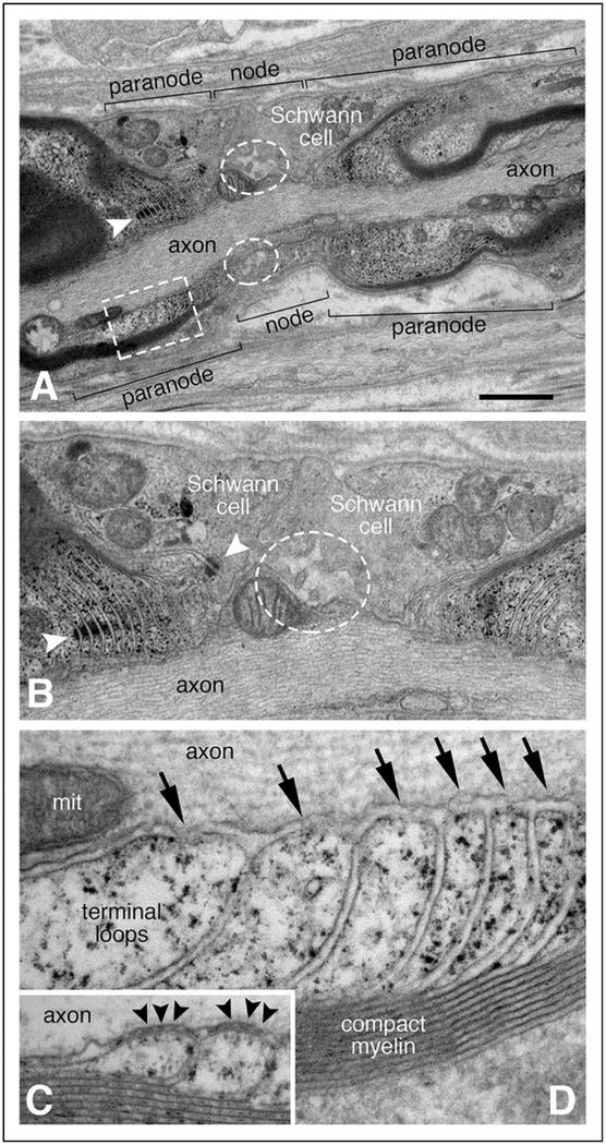
Abnormal node of Ranvier ultrastructure. Panels A, B, and D are from a single node of Ranvier observed in the sural nerve biopsy performed at 3.5 years of age; panel C is from a control adult sural nerve. The approximate borders for the node and the paranodal regions are delimited by brackets (A). Instead of normal microvilli, broad Schwann cell processes extend into the nodal region from either side (A and B). Only a small number of blunted microvilli are present at the node (white dashed line ovals in A and B). Normal appearing adherins junctions are noted between adjacent, uniform terminal loops of the left heminode (white arrowhead in A; left white arrowhead in B). The adherins junction nearest the node (right white arrowhead in B) marks the boundary of several abnormally shaped terminal loops. Many of the terminal loops (marked by the white dashed line rectangle in panel A and shown at higher magnification in D) appear to arise normally from compact myelin. However, the septate axoglial paranodal junctions that normally bridge between the tip of each terminal loop to the axolemma (black arrowheads in C) are absent from the patient (arrows in D). The size bar in panel A equals 1.2 μm in panel A, 0.6 μm in panel B, and 0.15 μm in panels C and D. The structure in the axoplasm of panel D labeled “mit” is a mitochondrion.

