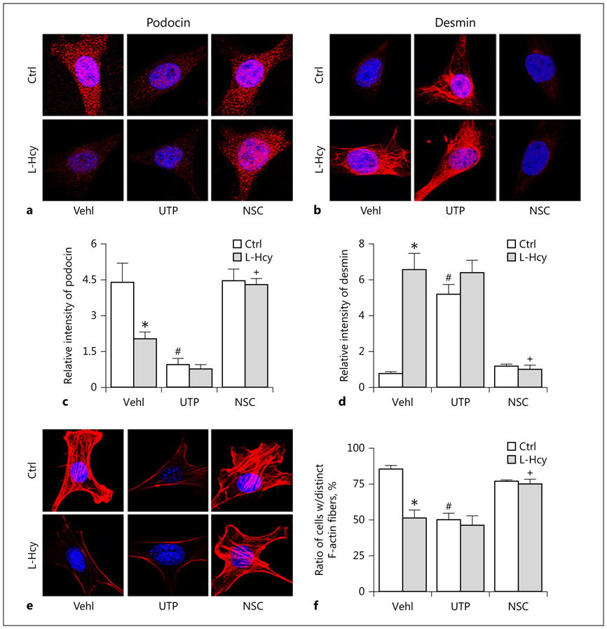
Fig. 3.
Attenuation of L-Hcy-induced podocyte dysfunction by Rac1 inhibition. a and b Immunofluorescence staining showed that activation of Vav2 by UTP or Rac1 inhibition by NSC rescued Hcy-induced expression of podocyte marker podocin as well as suppressed expression of podocyte injury marker desmin (n = 6). c and d Summarized graph showed the statistical data for podocin and desmin. e and f Microscopic images of F-actin proceeded rhodamine-phalloidin staining in podocytes and summarized data of distinct, longitudinal F-actin fibers. Scoring was from 100 podocytes in different groups (n = 6). * p < 0.05 versus Ctrl; # p < 0.05 versus Vehl/Ctrl; + p < 0.05 versus Vehl/L-Hcy. Vehl, vehicle; UTP, uridine triphosphate; NSC, NSC-23766; Hcy, homocysteine.

