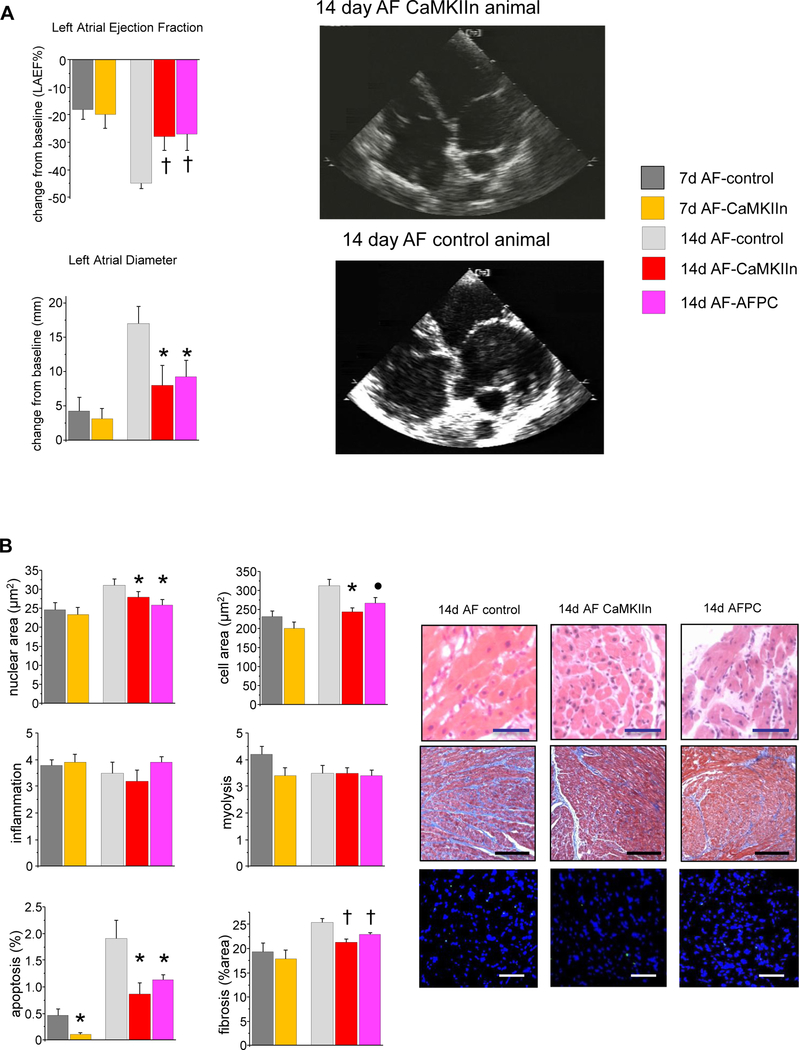
Figure 3.
Effects of CaMKII inhibition on structural remodeling. A: Echocardiography showed delayed progression in atrial dilation and dysfunction in subgroups receiving CaMKIIn. No change in ventricular structure or function was observed (data not shown). Right: Apical 4-chamber echocardiographic views illustrate the smaller atrial size in the CaMKIIn animals. B: Histological analyses show significantly less nuclear and cellular hypertrophy, apoptosis, and fibrosis in CaMKIIn-treated animals at 2 weeks, and decreased apoptosis at 1 week; otherwise, no significant changes were seen at that time point, and no effect of CaMKIIn gene transfer on myolysis or inflammation. Right: Representative images of right atrial microsections. Magnification bar indicates 50 μm in hematoxylin/eosin-and TUNEL-stained images and 200 μm in Masson trichrome images. ●P <.10; *P <.05; †P <.01. AF = atrial fibrillation; AFPC = atrial fibrillation pacing control.

