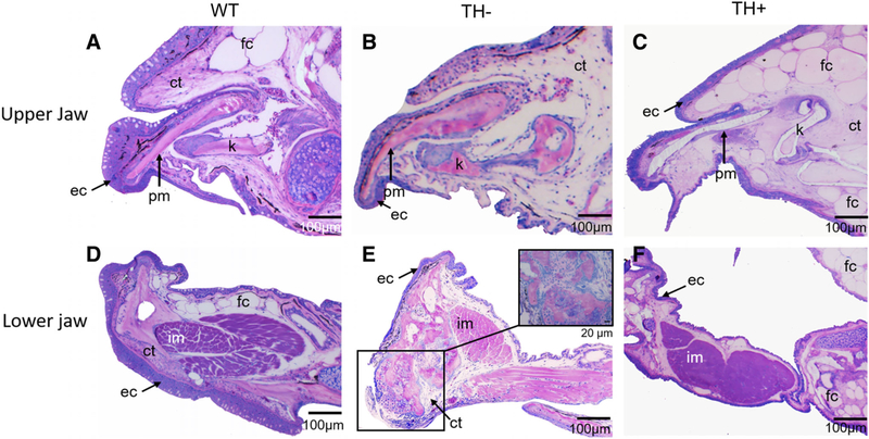
Fig. 6.
Parasagittal sections from WT, TH−, and TH+ zebrafish stained with Lee’s Methylene Blue-Basic Fuschin. (A, B, and C) Sections through upper jaw showing histological structure of premaxilla and kinethmoid. (D, E, and F) Sections through lower jaw showing histological structure of dentary and intermandibularis muscle. Note the thin epithelium, lack of fat cells, and disorganized connective tissue in the upper and lower jaw of TH−, and the thin epithelium, surplus of fat cells, and lack of connective tissue in TH+. In TH−, the intermandibularis muscle is smaller than in WT, while in TH+ it is larger. (E, inset) Closeup view of the endochondral bone growth at the tip of the dentary, present only in older TH− individuals.

