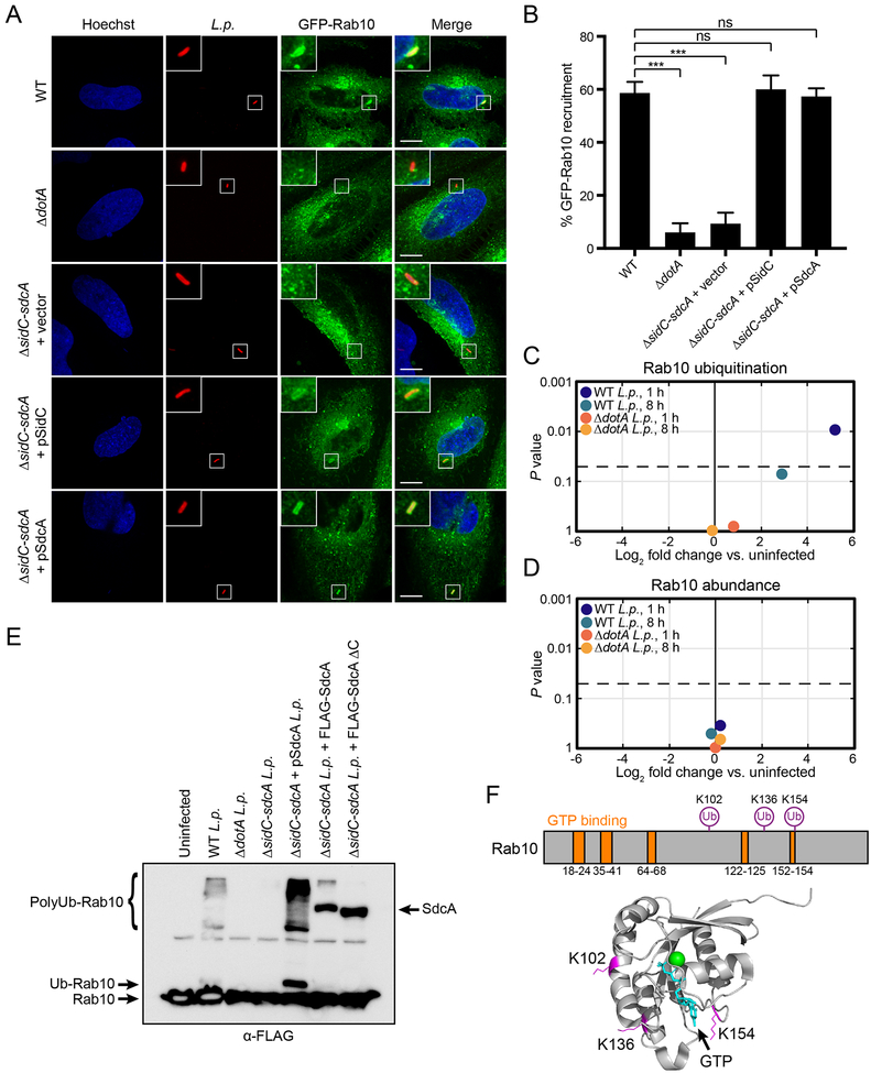
Figure 5. Rab10 is recruited to the LCV and ubiquitinated by SidC/SdcA during L. pneumophila infection.
(A) HeLa FcγRII cells stably expressing GFP-Rab10 were infected with WT, ΔdotA, or ΔsidC-sdcA complemented with empty vector, or plasmid-encoded SidC or SdcA L. pneumophila at MOI = 3 for 1 h, fixed, and immunostained for L. pneumophila (red). Nuclei were stained with Hoechst dye (blue). Scale bars, 10 μm.
(B) Quantification of the percentage of intracellular L. pneumophila co-localizing with GFP-Rab10 for the indicated L. pneumophila strains. Mutant L. pneumophila strains were compared to WT L. pneumophila (n = 3 technical replicates with at least 50 LCVs analyzed per replicate, two-tailed Student’s t test, ns = not significant, ***P<0.001; error bars, s.d.
(C and D) HEK293 FcγRII cells were infected with WT or ΔdotA L. pneumophila at MOI = 100 for 1 h or 8 h and Rab10 ubiquitination (C) and protein abundance (D) were analyzed by mass spectrometry (n = 3 biological replicates). Plots show log2 fold change vs. uninfected control (x-axis) vs. P value (y-axis). Dotted line represents p-value cutoff of 0.05.
(E) Immunoblot analysis of Rab10 ubiquitination. HEK293 FcγRII cells stably expressing 3xFLAG-Rab10 were either untransfected (lanes 1–5) or transfected with FLAG-tagged full-length SdcA or SdcA lacking residues 222–315 (lanes 6 and 7); 21 h after transfection, cells were either uninfected or infected with WT, ΔdotA, ΔsidC-sdcA, or ΔsidC-sdcA complemented with plasmid-encoded SdcA L. pneumophila. Cells were lysed 1 h post-infection and probed with anti-FLAG antibody.
(F) Schematic and 3D structure showing Rab10 residues ubiquitinated by L. pneumophila during infection. Green, Mg2+; Teal, GTP; Magenta, ubiquitinated residues.
See also Figure S5.

