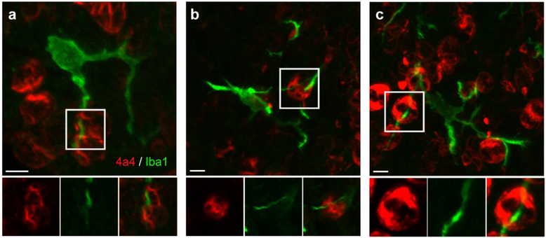
Figure 4.
The processes of some periventricular microglia are very closely affiliated with NPC soma and appeared to course through the soma of 4A4+ dividing NPCs. Panels A, B, and C show the projected images of periventricular microglia in the E19 rat. Insets below highlight processes from these cells that extended to the ventricle and appeared to course through the soma of mitotic NPCs. We interpret these images as microglial processes that course through grooves or channels in the outer membrane of the mitotic NPCs. Scale bars = 5 μm.

