Figure 5.
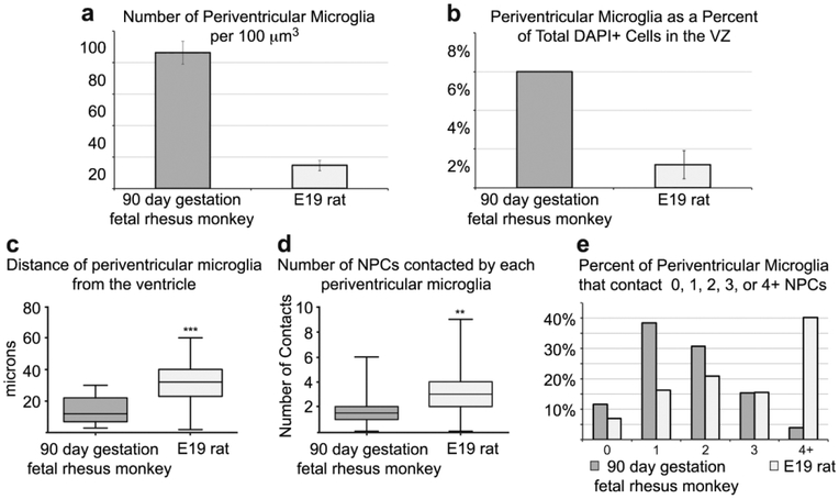
Comparison of periventricular microglia in the 90 days gestation fetal rhesus monkey and E19 rat. (A) The number of periventricular microglia is higher in the 90 days gestation fetal rhesus monkey compared to the E19 rat (p < 0.01,**). (B) The proportion of microglia in comparison to the total number of DAPI+ cells is higher in the periventricular zone of the fetal monkey. (C) Periventricular microglia were positioned significantly closer to the ventricle in 90 days gestation fetal rhesus monkey compared to E19 rat (p < 0.0001,***). (D) Periventricular microglia in the E19 rat contacted more NPCs that in the fetal rhesus monkey (p < 0.01,**). (E) Periventricular microglia in the E19 rat were more likely to contact more than one NPC. E19 rat and 90 days gestation rhesus monkey represent the same stage of cortical neurogenesis. Due to different NPC dynamics (Martinez-Cerdeño et al., 2012), fewer NPCs divide at the ventricle at this stage of development in the fetal rhesus monkey, the VZ is significantly thinner, and microglia may be positioned closer to the ventricle in the rhesus monkey as a result.

