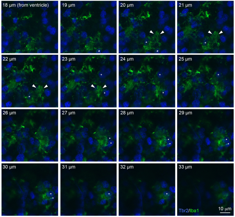
Figure 6.
Periventricular microglia (Iba1, green) in 90 days gestation fetal rhesus monkey had complex morphology and some of these cells extended large membranous sheets that contacted and enveloped neighboring Tbr2+ NPCs. The soma of this periventricular microglial cell was positioned 20 μm from the surface of the lateral ventricle and is indicated in optical planes 20-23 μm with white arrowheads. The cell extended a phagocytic cup that encircled a Tbr2+ NPC (hashtag) in optical planes 18-20 μm. Five Tbr2+ NPCs (blue) that were enveloped by the membranous extension from this periventricular microglial cell (asterisks) are visible in the optical planes at 20-22 μm (1 cell), 22-25 μm (2 cells) and 27-32 μm (2 cells). Scale bar = 10 μm. The Z-stack can be viewed as a QuickTime movie in Supplemental Movie 2.

