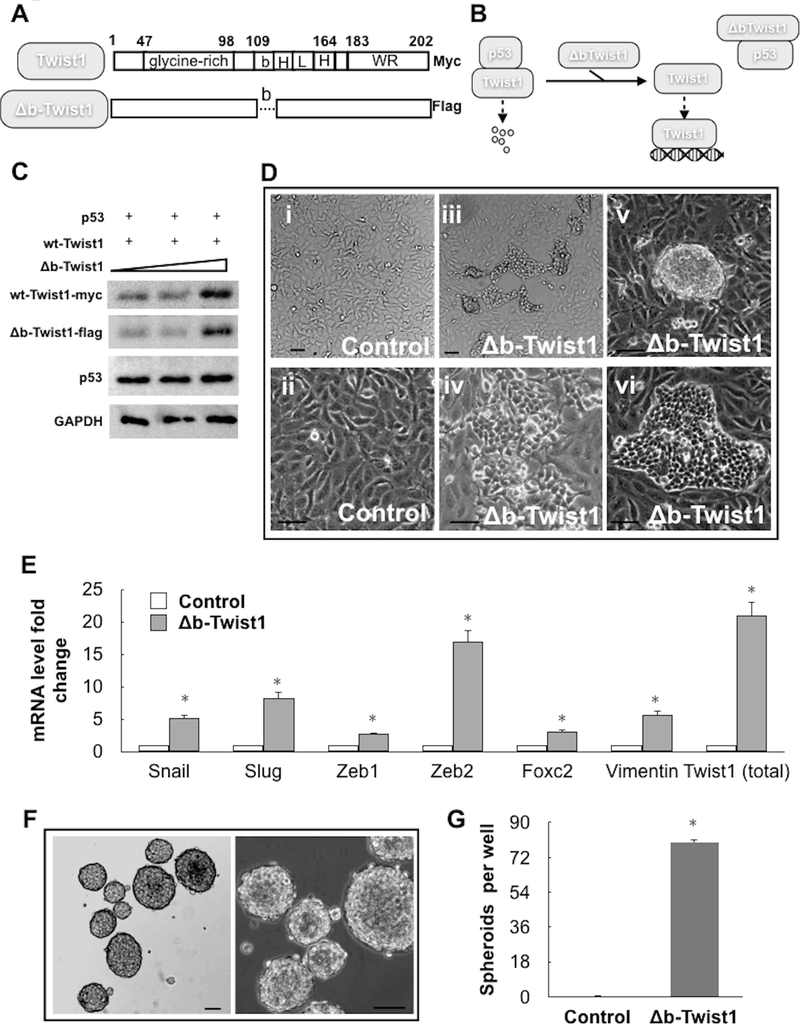
Figure 5. Inhibition of p53 and Twist1 interaction induces EMT in ovarian cancer cells.
(A) Schematic illustration showing the structure of the dominant negative Δb-Twist1 construct used compared to wild-type Twist1. Note difference in C-terminal tags. (B) Schematic illustration explaining how Δb-Twist1 competes with wild-type Twist1 to interact with p53, thus releasing and stabilizing wild-type Twist1. (C) HEK293T cells were transiently transfected with wild-type p53 and wild-type Twist1 in the presence of increasing concentrations of Δb-Twist1 and effect on protein expression determined by western blot. Anti-myc-tag and anti-flag-tag antibodies were used to distinguish wild-type Twist1 and Δb-Twist1, respectively. The amount of vectors are the same for each condition and equalized using empty-vector controls (D) Δb-Twist1 was stably overexpressed in R182 cells (p53-high/Twist1-) and its effect on morphology was determined by light microscopy. Control cells were transfected with empty-vector controls. Note that Δb-Twist1 is able to induce morphological changes associated with a fibroblastic/mesenchymal phenotype; i, iii, v taken at 4x; ii, iv, vi taken at 10x. (E) Expression of mesenchymal markers in samples from D was determined by QPCR; * = p<0.05 compared to empty-vector control. (G) R182 cells stably expressing Δb-Twist1 as in D were grown in ultra-low attachment plates and formed compact spheroid. (F) Quantification of spheroid formation in G. Data is presented as mean (+/− SEM). Note that the control cells transfected with empty vector did not form spheroids. *= p<0.05 compared to control. Representative of at least 3 independent experiments.

