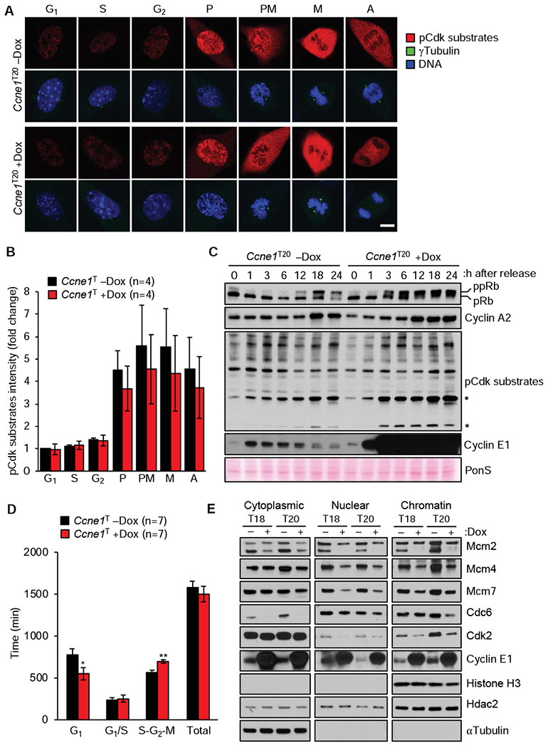
Figure 2.

High transgenic expression of cyclin E1 alters cell-cycle timing. (A) Representative images of dox-treated and untreated Ccne1T MEFs at the indicated stages of cell cycle immunolabeled for phosphorylated Cdk substrates. Scale bar, 5 μm. (B) Quantification of pCdk substrate signals of the indicated MEFs at various stages of the cell cycle (normalized to -dox G1, signals). (C) Western blots of lysates of Ccne1T MEFs harvested at the indicated time points after release from serum starvation in the presence or absence of dox. Asterisks mark hyperphosphorylated Cdk substrates. (D) Analysis of the indicated MEFs by FUCCI technology. (E) Western blots of fractionated lysates of MEFs grown with or without dox for 72 h. Histone H3, Hdac2, and αTubulin represent chromatin, nuclear and cytoplasmic markers, respectively. Data in B and D represent mean ± s.e.m. Statistics: B and D, two-tailed paired t-test. *P < 0.05, **P < 0.01.
