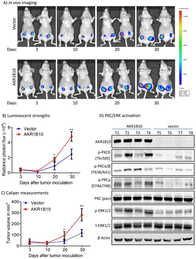
Figure 6. AKR1B10 promotes tumor growth in female nude mice.
MCF-7 cells (5 × 10⁶) labeled with luciferase were implanted in the mammary fat pads (orthotopic) of immunodeficient female mice at 4-6 weeks old. A pellet of 17β-estradiol (0.72mg/pellet) was implanted beneath the neck skin. (A) Representative in vivo bioluminescent images at days 3, 10, 20 and 30 post the cell injections. (B) Tumor volumes by in vivo bioluminescence at photon/second. (C) Caliper measurements of the tumor size. *, p<0.05 and **, p<0.01 when compared to vector control. (D) PKC/ERK signaling activity in tumors. *, p<0.05 and **, p<0.01 when compared to vector control.

