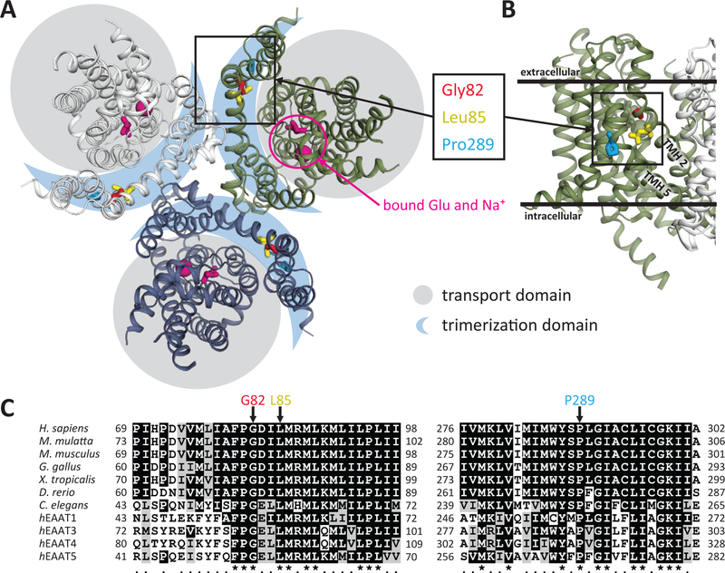
Figure 1. Structural relationship and evolutionary conservation of de novo SLC1A2 variants associated with epileptic encephalopathy.
(A) Crystal structure of thermostable SLC1A3 (PDB ID: 5LLU) marked using conserved residues homologous to SLC1A2 Gly82, Leu85 and Pro289 positions, and the glutamate substrate binding pocket.
(B) Close-up view of TMH2 and 5 in reference to the inner and outer layers of the cytoplasmic membrane as well as the trimerization domain.
(C) ClustalW 2.1 sequence alignment of SLC1A2 orthologs and different EAAT proteins for the SLC1A2 protein regions containing G82, L85 and P289.

