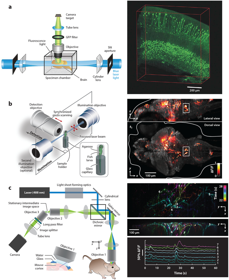
Figure 2.
Examples of light-sheet approaches for neuroscience applications. (a) Demonstration of ultramicroscopy for light-sheet imaging of cleared samples (left), including green fluorescent protein (GFP)-expressing neurons in the mouse brain (right). Panel a adapted with permission from Dodt et al. (2007). (b) Whole-brain imaging of calcium activity (GCaMP) in a zebrafish larva at around 1 volume per second (right). (Left) The system used scanned light sheets incident from both sides of the sample, with the detection lens moved by a piezo actuator, to maintain focus on the sheet. The fish was positioned in an agarose tube between the three objective lenses. Panel b adapted with permission from Ahrens et al. (2013). (c) Swept confocally aligned planar excitation (SCAPE) imaging of GCaMP activity in apical dendrites of layer 5 neurons in awake, behaving mouse cortex at 10 volumes per second. (Right, top and middle) Time-color-encoded firing events over a 6-s period shown as maximum-intensity projections over z and x. (Right, bottom) Raw time courses of fluorescence change (ΔF/F) extracted from the locations indicated in the right, top and middle panels, demonstrating minimal photobleaching and a high signal-to-noise ratio. Panel c adapted with permission from Hillman et al. (2018).

