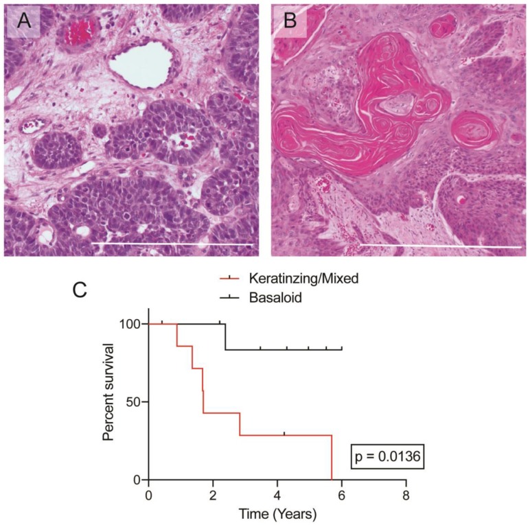
Figure 1. Histologic images of anal squamous cell carcinoma.
(A) Representative image of a “basaloid” architecture in ASCC, 100X. (B) Representative image of a “keratinizing” architecture in ASCC, 100X. (C) Kaplan–Meier curves plotting overall survival based on histologic architecture. Bar = 500 μm.

