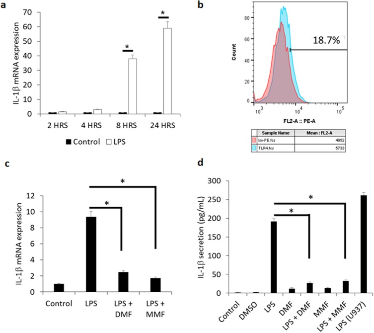
Figure 1.
DMF and MMF inhibit the production and release of LPS-mediated IL-1β release from NK92 cells. (A) Time-dependent treatment of NK92 cells with 10 μg/mL LPS upregulates the expression of IL-1β mRNA. *P values (<0.01) compare the mRNA expression in LPS-activated cells (white columns) versus the background control (black columns). (B) Flow cytometry showing representative experiment of the binding of isotype control antibody (red color) and antibody against TLR4 (blue color). Percentages of positive cells are shown. (C) Comparison of mRNA expression of IL-1β among LPS-stimulated cells and those treated with LPS plus DMF or MMF. *P<0.01. (D) LPS-induced IL-1β release and its inhibition by DMF or MMF, as detected by ELISA assay. NK92 cells were incubated with 10 μg/mL LPS either alone or in the presence of 100 μM DMF or MMF in media containing 200 IU/mL IL-2 for 24 h. *P values (<0.01) compare IL-1β release in LPS-activated cells vs cells incubated with LPS plus DMF or MMF. U937 cells treated with 10 μg/mL LPS for 24 h were used as a positive control in these experiments.

