
Figure 5.
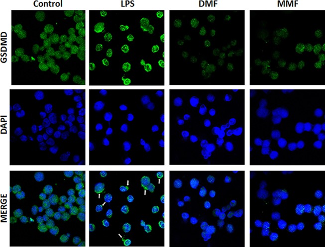
Confocal microscopyimages showing the expression and translocation of GSDMD. NK92 cells were either left untreated (Control) or incubated with 10 μg/mL LPS in the absence or presence of 100 μM of either DMF or MMF, and in the presence of IL-2. DPI was used to stain the nuclei. Arrows indicate pyroptotic bodies after treatment of NK92 cells with LPS.