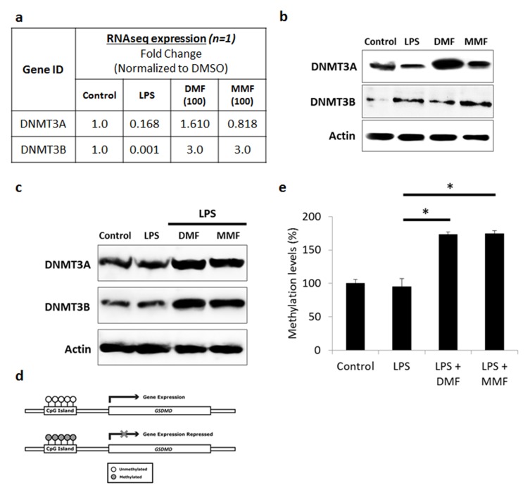
Figure 6.
DMF and MMF induced DNMT-mediated methylation-silencing of GSDMD gene. (A) Whole transcriptome analysis data showed increase in DNMT3A and DNMT3B in DMF- or MMF-treated NK92 cells, compared to control cells. (B) Western blotting data also showed an increase in the expression of DNMT3A and DNMT3B in DMF or MMF treated cells but not in LPS treated cells. (C) DMF and MMF increased the expression of DNMT3A and DNMT3B in cells incubated with LPS. (D) Schematic representation of the presence of CpG Islands in the promoter region of GSDMD gene and repression of expression is observed upon methylation of those CGIs. (E) Data from MSP-PCR showed the methylation levels of GSDMD in cells either untreated (Control), or treated with LPS, or LPS plus DMF or MMF in the presence of IL-2 for 24 h. *P<0.01 compares GSDMD promoter methylation levels in LPS-activated cells vs cells incubated with DMF or MMF. Methylation levels were significantly higher in DMF and MMF treated NK92 cells compared to untreated or LPS treated NK92 cells.

