Fig. 3.
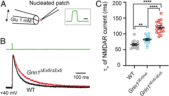
Deletion of Grin1 exon 5 results in a slower NMDAR deactivation. (A) Schematic view of the experimental setup. Brief pulses (2 ms) of l-glutamate (Glu, 1 mM) were applied to nucleated patch using a piezo-electrical device. NBQX (10 μM) and glycine (100 μM) were present continuously. The insert on the right shows the speed of solution exchange when switching from ACSF to 30% ACSF. The 20–80% time was 117 μs for the onset and 108 μs for the offset. (Scale bar, 2 ms.) (B) Normalized NMDAR-mediated currents in response to 2-ms glutamate pulse recorded from nucleated patches obtained from a WT and a Grin1ΔEx5/ΔEx5 neuron at P14. The green trace on the top indicates the pulse application of glutamate. Recordings were performed at room temperature (21–23 °C). (C) Decay constants of NMDAR-mediated currents recorded from WT (14 cells from 3 mice), heterozygous (15 cells from 3 mice), and homozygous mutant neurons (17 cells from 4 mice) at P14-15. ****P < 0.0001, **P < 0.005, Mann–Whitney U test.

