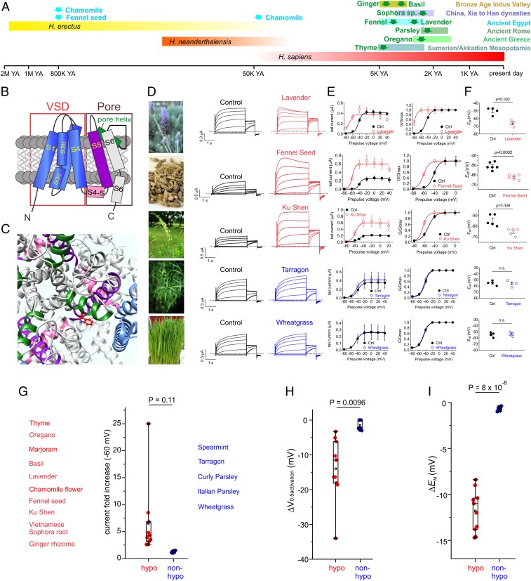
Fig. 1.
KCNQ5 activation is a specific, shared feature of botanical hypotensive folk medicines. All error bars indicate SEM. (A) Approximate timeline of human use of the hypotensive plants examined in this study. YA, years ago. (B) Topological representation of KCNQ5 showing 1 of the 4 subunits that comprise a channel. VSD, voltage sensing domain. (C) Extracellular view of the chimeric KCNQ1/KCNQ5 structural model to highlight the anticonvulsant binding pocket (red; KCNQ5-W235) using the color coding as in B. (D) A subset of the plants screened in this study (SI Appendix, Fig. S2 shows full screening data). (Left) Images of the plants used (attributions provided in SI Appendix). (Right) Mean TEVC current traces for KCNQ5 expressed in Xenopus oocytes in the absence (control) or presence of 1% extract from the medicinal plants as indicated (n = 4 to 5). Dashed line here and throughout indicates zero current level. Red indicates plants previously reported to show hypotensive activity or used traditionally as hypotensives; blue indicates nonhypotensives or diuretic hypotensives (here and E–I). (E) Mean tail current (Left) and normalized tail currents (G/Gmax; Right) versus prepulse voltage relationships for the traces as in D (n = 4 to 5). (F) Effects of the hypotensive plant extracts shown in D on resting membrane potential (EM) of unclamped oocytes expressing KCNQ5 (n = 4 to 5). (G–I) Scatter plots showing mean effects for the full screen (SI Appendix, Fig. S2) of hypotensive (red) versus nonhypotensive (blue) 1% plant extracts on (G) current at −60 mV, (H) V0.5activation, and (I) EM in oocytes expressing KCNQ5 (n = 4 to 8). Each point represents the mean data from 1 plant species.

