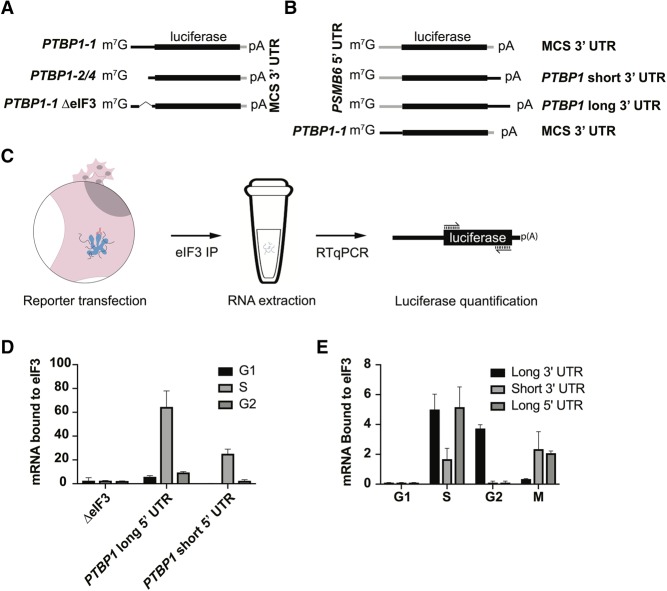
FIGURE 7.
Differential binding of PTBP1 UTR elements to eIF3 across the cell cycle. (A,B) Schematics of the luciferase reporters used in the experiments. (C) Schematic of the transfection, immunoprecipitation, and quantification method used to determine luciferase reporter mRNA binding to eIF3. (D) Distribution of binding to eIF3 across the cell cycle for the different PTBP1 5′-UTR elements as well as the deletion mutant. (E) Distribution of binding to eIF3 across the cell cycle for the different PTBP1 3′-UTR elements, as well as the long form of the PTBP1 5′-UTR. Binding experiments were carried out in biological triplicate, with standard deviation shown. Luciferase arbitrary units were normalized to WT for graphing.

