Fig. 1.
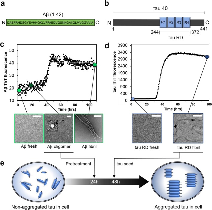
Characterization of Aβ and tau self-assembly. a The sequence of the Aβ(1–42). b A schematic of full-length tau (tau40, residue 1–441), which contains four tandem repeats (R1–R4) in the repeat domain (tau RD, residues 244–372). c ThT fluorescence (upper panel) and EM images (lower panel) of Aβ42 at different incubation times demonstrate that freshly prepared Aβ42 self-assembles and aggregates into spherical oligomers at 18 h and unbranched fibrils at 100 h. The inset highlights the spherical shape of the Aβ42 oligomers. The scales bar denotes 200 nm. d ThT fluorescence (upper panel) and EM images (lower panel) of tau RD. e A schematic representation of the timeline used for seeding experiment. Different Aβ42 self-assembly states and tau seeds were sequentially added to cells at 24 h and 48 h, respectively

