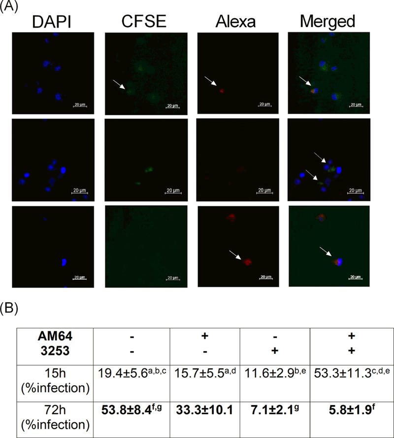
Figure 2: Percentage of human monocytes single- and co-infected with trypomastigotes from AM64 and 3253 strains.

Adherent cells obtained by plating PBMC for 1h (Panel A) or total human blood (Panel B) from non-Chagas donors were co-cultured with AM64 or 3254 trypomastigotes previously labeled with Alexa-Fluo or CFSE, respectively, as described in material and methods. (A) shows representative confocal microscopy analysis, showing double infected monocytes (first row, last figure - merged), CFSE-labeled 3253 single-infected (second row) and Alexa-labeled AM64 single-infected (third row) monocytes after 72 hours of infection. Arrows indicate monocytes co-infected with AM64 and 3253 (first row), single infected with 3253 (second row) and single infected with AM64 (third row); (B) Percentage of infection after 15 or 72 hours of culture obtained in whole blood incubated with both strains and acquired by flow cytometry. Results are expressed as average ± standard deviation. Identical letters indicate statistical significance, a: p= 0.04, b: p= 0.043, c: p= 0.007, d: p= 0.003, e: p= 0.005, g: p= 0.036 and f: p= 0.008.
