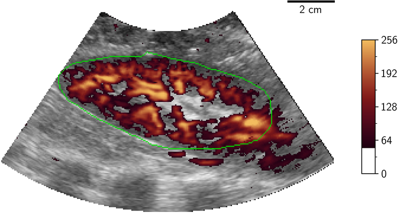
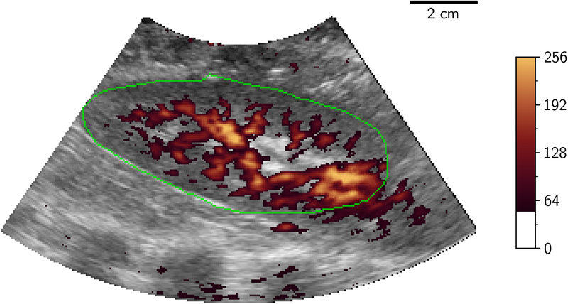
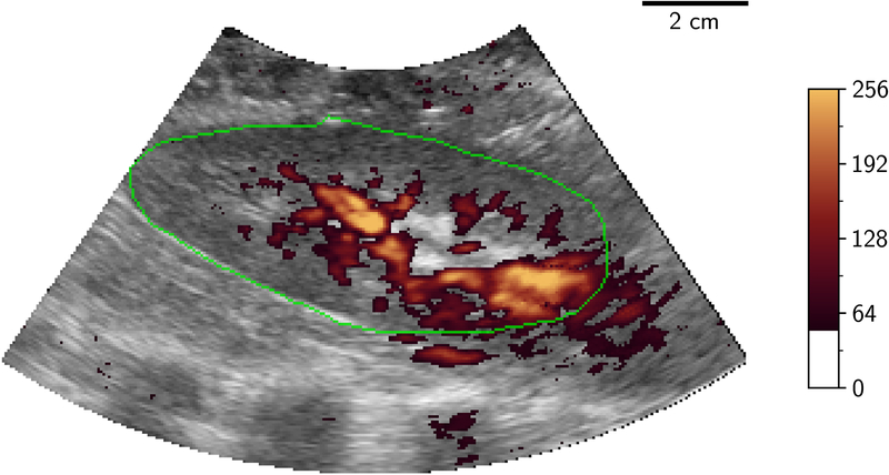
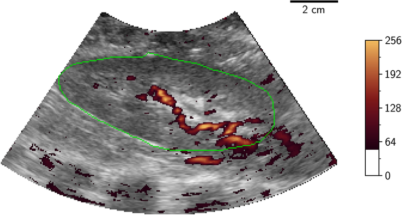
Figure 2.
2D B-mode (greyscale) slice of 3D-US volume with power Doppler mapping (orange) of porcine kidney demonstrating perfusion at different flow conditions: 100%, 75%, 50% and 25% (A to D). The renal outline is highlighted with the green line. 2D two dimensional; 3D three dimensional; US ultrasound.


