. Author manuscript; available in PMC: 2020 Oct 17.
Published in final edited form as: Cell Chem Biol. 2019 Aug 15;26(10):1365–1379.e22. doi: 10.1016/j.chembiol.2019.07.013

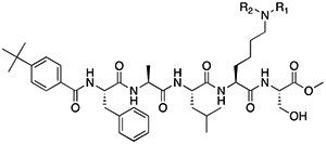
Table 1.

Methyl Lysine (Kme) Mimetic Compounds Screened in Polycomb in-vivo Assay.

graphic file with name nihms-1537978-t0002.jpg
Kme Mimetic EC50 (μM)
graphic file with name nihms-1537978-t0003.jpg 105.1 ± 11.25
graphic file with name nihms-1537978-t0004.jpg 41.66 ± 2.240
graphic file with name nihms-1537978-t0005.jpg 36.57 ± 1.140
graphic file with name nihms-1537978-t0006.jpg 26.55 ± 3.240
graphic file with name nihms-1537978-t0007.jpg 13.74 ± 0.535
graphic file with name nihms-1537978-t0008.jpg 33.08 ± 1.905
graphic file with name nihms-1537978-t0009.jpg 23.73 ± 1.740
graphic file with name nihms-1537978-t0010.jpg ~ 20
graphic file with name nihms-1537978-t0011.jpg 4.590 ± 0.292
graphic file with name nihms-1537978-t0012.jpg 3.207 ± 0.352
graphic file with name nihms-1537978-t0013.jpg 1.018 ± 0.175
graphic file with name nihms-1537978-t0014.jpg 2.356 ± 0.115
graphic file with name nihms-1537978-t0015.jpg 2.223 ± 0.194
graphic file with name nihms-1537978-t0016.jpg 2.057 ± 0.135

Peptidomimetic scaffold shown above table for reference, with methyl-lysine mimetic modification site highlighted in blue. Data are presented as mean ± SD from three biological replicate experiments.