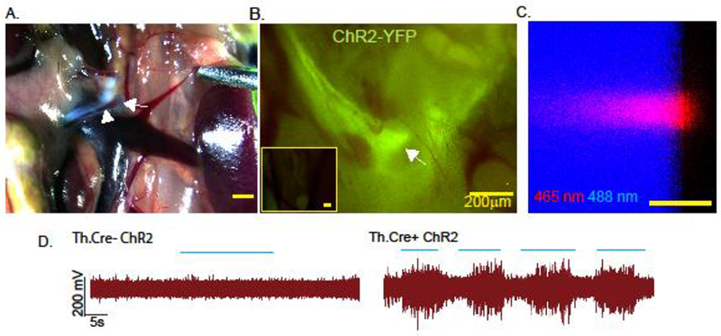
Fig. 1. Site specific peripheral neuronal activation by optogenetics.

The superior mesenteric ganglion (arrow) was identified using the superior mesenteric nerve (arrowheads) as an anatomical landmark under a brightfield stereodissection microscope (A). YFP expression was detected in Th.Cre+ ChR2-YFPLSL but not Th.Cre- ChR2-YFPLSL mice (inset) was assessed using a stereo-epifluorescence microscope with a YFP bandpass filter (B). Characterization of the size of light emitted from the fiberoptic patch cable by confocal microscopy using on edge illumination of a plexiglass slide. Boundaries of the slide are revealed using 488 nm laser light excitation from the microscope (C). Optogenetic stimulation (blue lines indicate “pulses of light”) of the SMG results in compound action potentials in the superior mesenteric nerve of Th.cre+ ChR2 but not Th.Cre- ChR2 mice (D). Images and electrical recordings are representative of 3–4 mice, or 3 separate experiments in the case of “B”.
