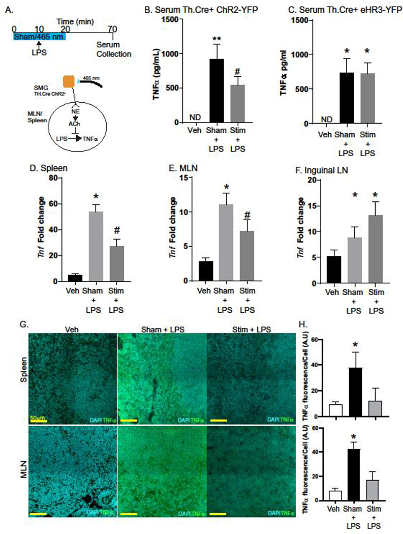
Fig. 2. Activation of sympathetic neurons in the SMG blocks LPS-induced TNFα production.

To test the ability of sympathetic neurons in the SMG to regulate LPS-induced TNFα production, a conditional optogenetic approach was used. After exposure of the SMG, mice were either subjected to sham (no light) or 465 nm light pulses for 10 minutes prior to i.v. injection of LPS (4 mg/kg) and continued for a total of 20 minutes (Stim). Vehicle control mice received sterile PBS (i.v.) (A). Serum collected revealed LPS induced TNFα production was significantly lower in stimulated (ChR2) vs. Sham treated mice (B). Mice that express the inhibitory halorhodopsin-YFP did not exhibit 465 nm induced reductions in TNFα production (C). Expression of TNFα mRNA was significantly reduced in ChR2 stimulated LPS treated mice compared to Sham in the spleen (D) and MLN (E) but not the inguinal LN (F). Tissues from these mice were evaluated by confocal microscopy (G) to determine TNFα production by fluorescence per DAPI+ cell (H). n= 5–8 mice/group in 2 experiments with #, * P<0.05 ANOVA Tukey post-test compared to all groups. ND= none detected, data are presented as mean ± SEM for all panels. qPCR results are fold expression normalized to control and the endogenous Actb gene as described in the methods.
