
Fig 4. Sympathetic innervation is sparse in the MLN.
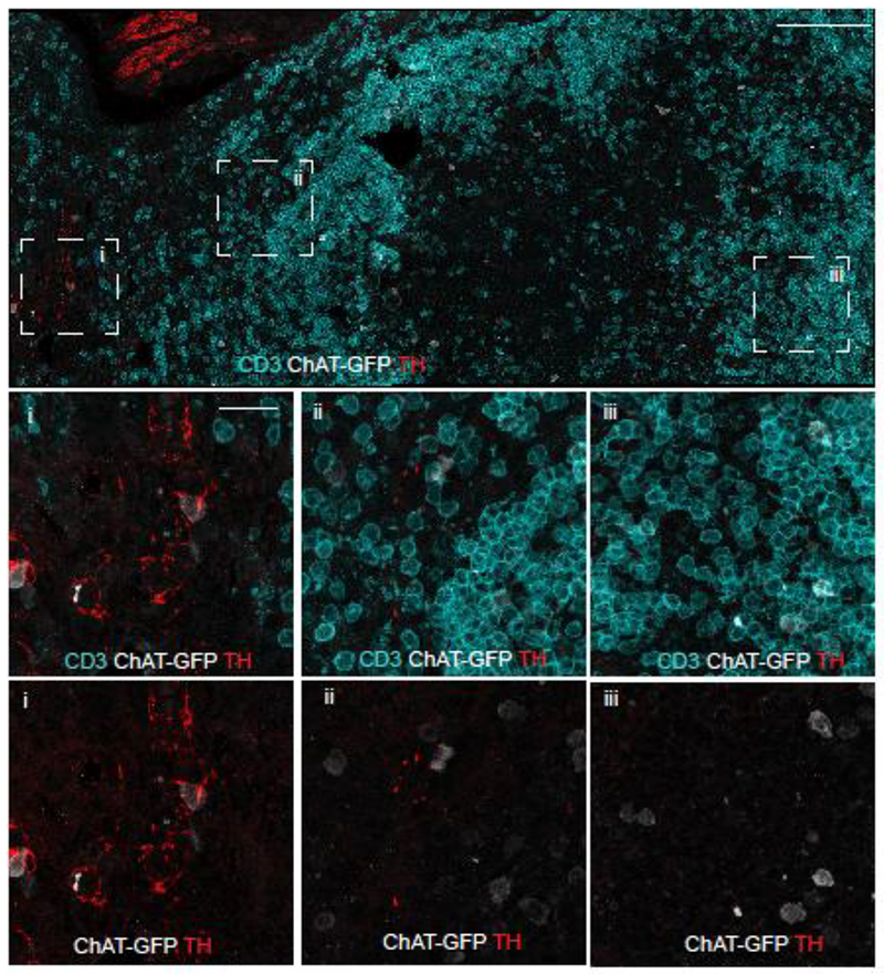
Confocal microscopy was conducted on tissue sections ChAT-GFP (C57BL/6 background) mice for T-cells (anti-CD3), ChAT (anti-GFP), and sympathetic axons (anti-TH). The section was identified and overlapping tiles were acquired from 943 µm x 454 µm area of MLN (upper panel, scale bar =100 µm). Areas indicated in the bounding boxes are shown in the middle (CD3, ChAT-GFP, TH) and lower panels (ChAT-GFP, TH, scale bar = 20 μm). Region of the hilus in the MLN is within box “i”. Representative images obtained from 8 mice.