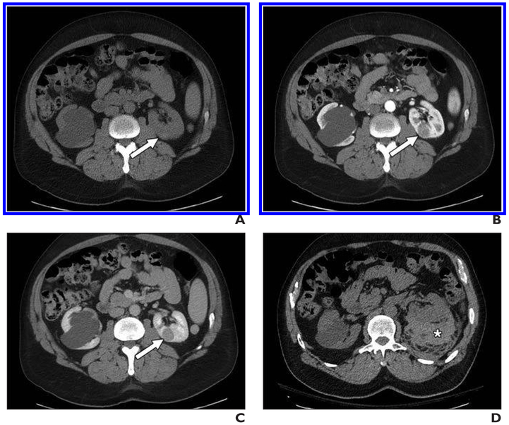
Fig. 1. 56-year-old man with left renal mass.
A–C, Unenhanced axial CT image (A) and contrast-enhanced axial CT images acquired during corticomedullary (B) and nephrographic (C) phases show homogeneous and progressively enhancing mass in posterior medial left kidney (arrow), which is mildly hyperattenuating on unenhanced image. Mass was favored to represent papillary renal cell carcinoma (pRCC), and patient underwent percutaneous renal mass biopsy.
D, Postbiopsy CT was performed because patient reported pain. Representative unenhanced CT image shows moderate perinephric hematoma along posterior left kidney (asterisk). Patient was admitted to hospital and observed; no transfusion or other support other than observation was reguired. Biopsy confirmed diagnosis of pRCC. After partial nephrectomy, final diagnosis of type I pRCC was rendered.

