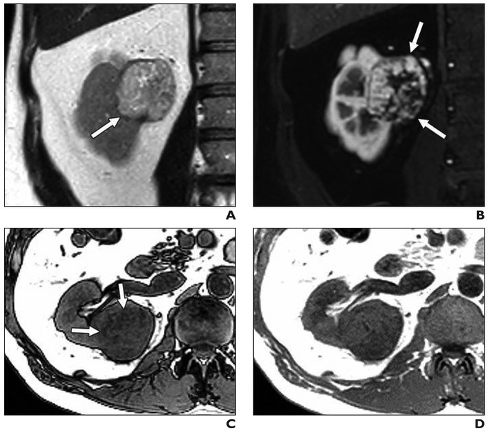
Fig. 5. 47-year-old man with renal mass.
A, Coronal T2-weighted single-shot fast spin-echo image shows mass in medial right kidney (arrow) that is heterogeneously hyperintense relative to renal cortex.
B, On coronal fat-saturated T1-weighted gradient-echo image acquired during corticomedullary phase, lesion (arrows) shows intense heterogeneous enhancement.
C and D, Region of decreased signal intensity (arrows, C) is present on axial opposed-phased gradient-echo T1-weighted image compared with in-phase gradient-echo image (D); this finding is consistent with microscopic fat within tumor. This lesion was given clear cell likelihood score of 5. Patient underwent partial nephrectomy, and histopathology showed clear cell renal cell carcinoma, International Society of Urological Pathology grade 3.

