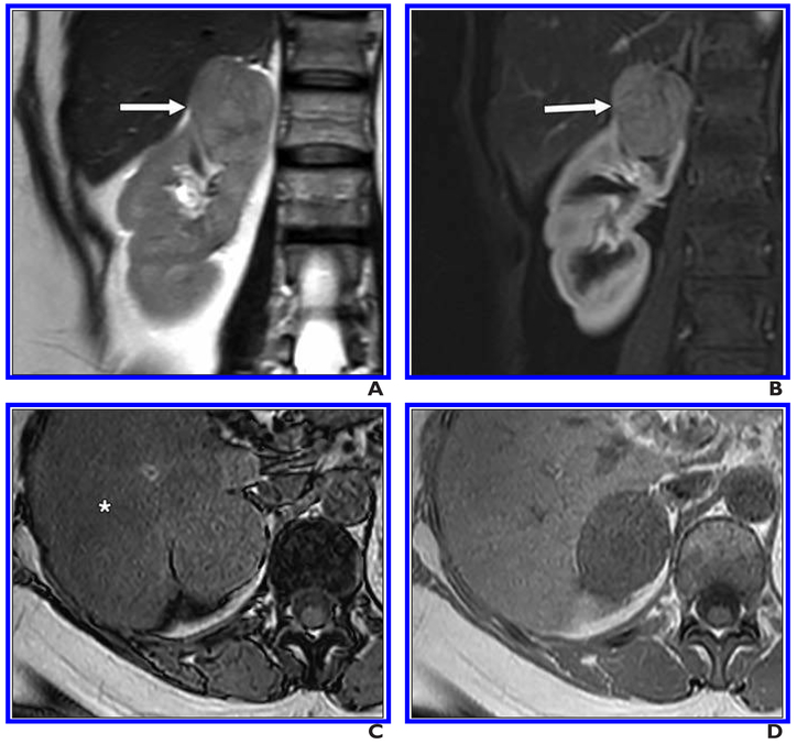
Fig. 7. 44-year-old man with solid right renal mass.
A, Coronal T2-weighted single-shot fast spin-echo image shows mildly heterogeneous mass (arrow) is isointense to renal cortex and arising from upper pole of right kidney.
B, Coronal fat-saturated T1-weighted gradient-echo image acquired during corticomedullary phase shows moderate enhancement in mass (arrow).
C and D, Axial opposed-phase (C) and in-phase (D) gradient-echo T1-weighted images show no intralesional fat. Incidentally, decreased signal intensity (asterisk, C) in liver consistent with hepatic steatosis is noted on opposed-phase imaging. This lesion was given clear cell likelihood score of 3. Patient underwent percutaneous renal mass biopsy, and histopathology revealed chromophobe renal cell carcinoma.

