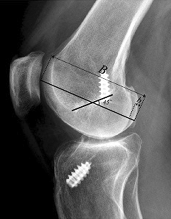
Fig. 3.

The radiograph shows the newly developed femoral condyle overlap method, used to validate rotation for lateral radiographs. A line was drawn from the posterior to the anterior edge of the femoral condyle (B), intersecting the midpoint at a 45° angle. The angle size was determined as 45°, since it was estimated to represent the anteroposterior axis of the femoral condyle. Moreover, a line (b) was drawn from the posterior limit of the femoral condyle to the border of the femoral condyle situated just anteriorly (corresponding to the medial or lateral condyle depending on the direction of the rotation of the knee), whereafter the femoral condyle overlap can be calculated using an equation
