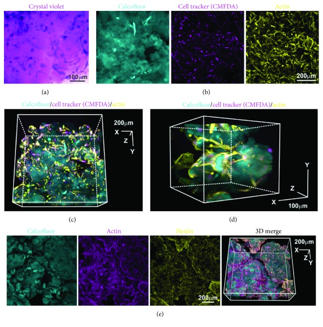
Figure 5.
aNFC is compatible with multiple light microscopy-based assays. (a) ADSCs were cultivated within aNFC and stained using crystal violet. Bright-field microscopy revealed evenly distributed, dark-stained ADSCs. Bar: 100 μm. (b) ADSCs embedded in 0.2% aNFC were stained using cell tracker CMFDA (magenta) and phalloidin (yellow). aNFC was counterstained with calcofluor. A maximum intensity projection is shown. Bar: 200 μm. (c, d) 3D reconstruction demonstrated even staining intensity in all dimensions of the aNFC hydrogel. Bar in (c): 200 μm; bar in (d): 100 μm. (e) Immunocytochemistry with subsequent confocal microscopy was applied to visualise actin and the intermediate filament nestin in ADSCs in 0.2% aNFC. Bar: 200 μm.

