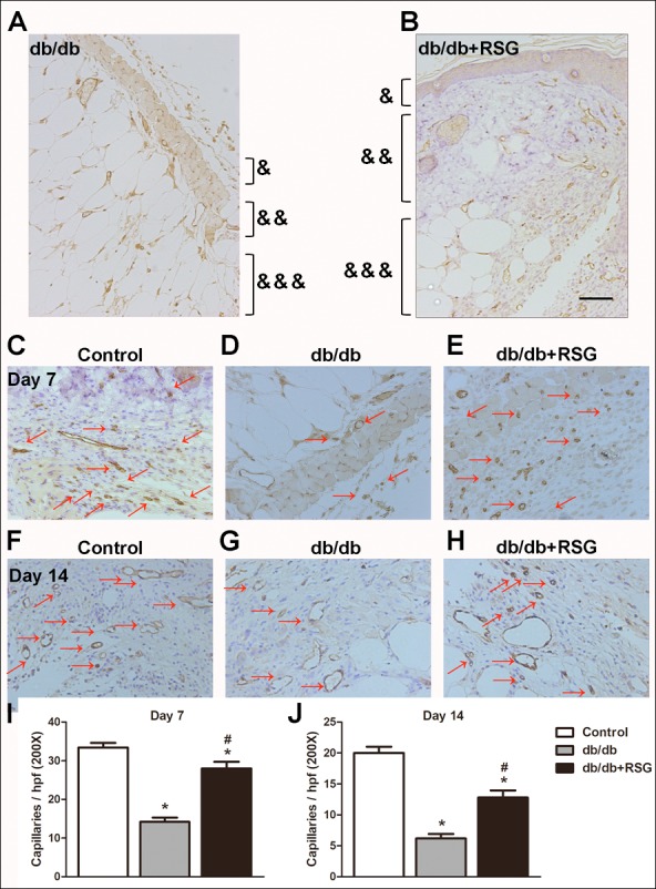
Figure 3. Rosiglitazone administration increased wound angiogenesis in db/db diabetic mice.

In db/db mice, after wound closures were made, skins around the wound were collected and angiogenesis was evaluated on days 7 and 14. Representative pictures of tissue destruction of wounds (A, B) and CD31 staining (C-H). Quantitative study on day 7 (I) and day 14 (J). Red arrows point out CD31-positive capillaries (200 ×; scale bar, 50 µm). Values are mean ±SEM, (n = 5 per group). ∗P < 0.05 vs. Control; #P < 0.05 vs. db/db. (&) Epidermis, (&&) dermis and (&&&) subcutis indicated. RSG, rosiglitazone. hpf, high power field.
