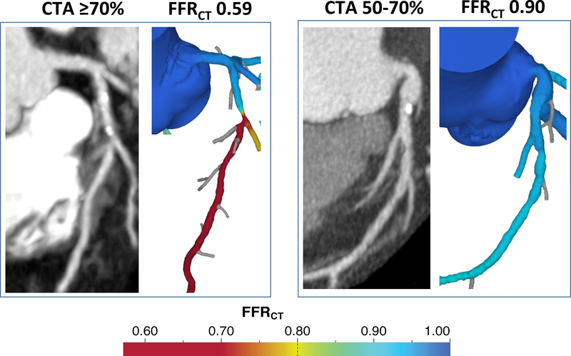
Figure 2.
Representative cases of coronary CTA and FFRCT. Left panel: 64-year-old man with acute chest pain diagnosed with unstable angina. Coronary CTA demonstrated 70–99% stenosis of the left anterior descending coronary artery at the origin of the first diagonal branch. FFRCT was abnormal (0.59). Invasive coronary angiography confirmed ≥70% stenosis and patient underwent stent placement. Right panel: 61-year-old woman diagnosed with non-cardiac chest pain. Coronary CTA demonstrated 50–70% stenosis in mid left anterior descending coronary artery. FFRCT was normal (0.90). Invasive coronary angiography showed 30–50% stenosis.

