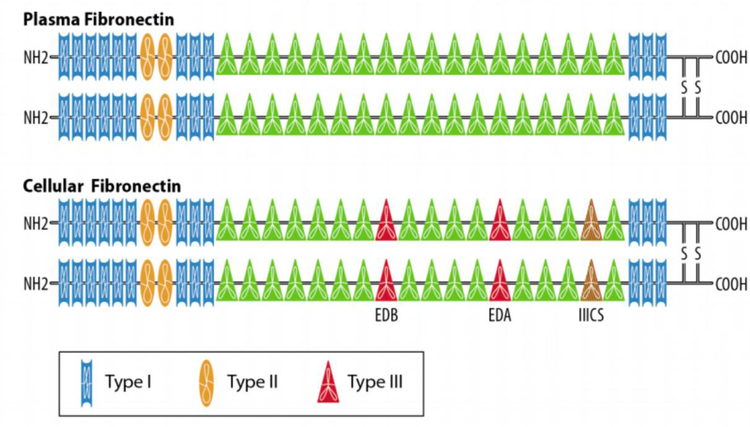
Figure 1.
Splice Variation Produces Differences in Fibronectin. Cellular fibronectin (bottom) differs from plasma fibronectin (top) by a number of additional repeats. Both types of fibronectin have 12 Type I repeats (rectangles) and 2 Type II repeats (ovals). Plasma fibronectin has 15 Type III repeats and cellular fibronectin typically has 17 Type III repeats (triangles). The IIICS splice site can introduce partial Type III repeats as well. EDA sits between the 11th and 12th Type III repeat and EDB sits between the 7th and 8th Type III repeat. These extra domains allow cellular fibronectin to interact with various integrin heterodimers.

