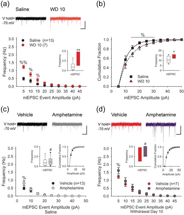
FIGURE 4.
Amphetamine enhances spontaneous mEPSCs. (a) Representative traces (above) show mEPSCs recorded in SPNs from saline- (left) and amphetamine-treated mice on WD 10 (right). The average frequency (inset) and the cumulative frequency distribution of mEPSCs was greater in withdrawal. For panels a and b: *p<0.05, **p<0.01, 2-tailed Student’s t-test; %p<0.05, %%p<0.01, 2-tailed Student’s t-test with Bonferroni correction. Bars: 10 pA, 1 sec. (b) The average mEPSC amplitude is greater on WD 10 (inset) and the cumulative amplitude distribution shows a greater fraction of mEPSCs in the 15-30 pA range. (c) Representative traces (above) show mEPSCs in SPNs from saline-treated mice in vehicle (upper left) and 5-7.5 min after bath-applied amphetamine (upper right). Amphetamine (10 μM) in vitro enhanced the frequency of mEPSCs (inset, left) by increasing the number of high-frequency, low-amplitude spontaneous inward currents but had no effect on the cumulative mEPSC amplitude distribution (inset, right). For panels c and d, #p<0.05, paired t-test. %p<0.05, %p<0.05, paired t-test with Bonferroni correction. (d) Representative traces (above) show mEPSCs in SPNs from amphetamine-treated mice on WD 10 in vehicle (upper left) and 5-7.5 min after bath-applied amphetamine (upper right). On WD 10, amphetamine (10 μM) in vitro boosted the frequency of mEPSCs (inset, left) by increasing high-frequency, low-amplitude inward currents, while having no effect on the cumulative amplitude distribution (inset, right).

