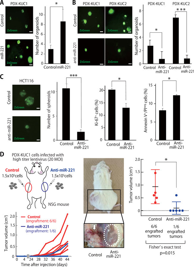
Figure 2. Inhibition of miR-221 reduces both the in vitro three-dimensional (3D) organoid-forming capacity and in vivo tumorigenicity of human CRC cells.
(A-B) Representative appearance and number of organoids formed by colorectal cancer PDX cells following infection with lentivirus vectors encoding either miR-221 (A) or an anti-miR-221 construct (B) (n=5; *p<0.05, ***p<0.001). scale bar: 100 μm. (C) Infection of HCT116 cells with a lentivirus vector driving constitutive miR-221 expression was associated with a reduction in 3D spheroid forming capacity (n=3; ***p<0.001), a reduction in the percentage of Ki67+ cells (n=3; *p<0.05), and an increase of the percentage of Annexin-V+/Propidium Iodideneg (PIneg) cells (n=3; *p<0.05). (D) Schematic illustration of in vivo xenotransplantation experiments and growth curves of tumors originated from PDX-KUC1 cells infected with either a lentivirus vector encoding for the anti-miR-221 construct or an empty vector used as negative control (n=6; 1.5×10⁵ cells/injection). Two months after xenotransplantation, PDX-KUC1 cells infected with the control vector formed tumors in 6 out 6 cases (100%), while those with the anti-miR-221 construct formed tumors in only 1 out 6 cases (17%; *p<0.05). All sub-cutaneous injection sites were dissected and visually inspected.

