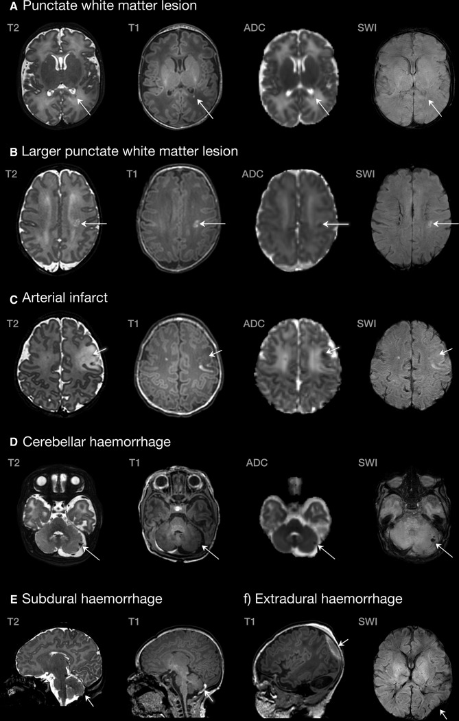
Figure 1.
Examples of lesions identified in the congenital heart disease cohort. (A) Single lesion in the posterior periventricular white matter (TGA, scanned at 39+6); (B) larger white matter lesion in the centrum semiovale (pulmonary atresia, scanned at 37+2); (C) left middle cerebral artery infarct (TGA, scanned at 39+5); (D) cerebellar haemorrhage (CoA, scanned at 39+3); (E) subdural haemorrhage (TGA, scanned at 39+2); (F) extradural haemorrhage (CoA, scanned 39+3). ADC, apparent diffusion coefficient; CoA, coarctation of the aorta; SWI, susceptibility-weighted imaging; TGA, transposition of the great arteries.

