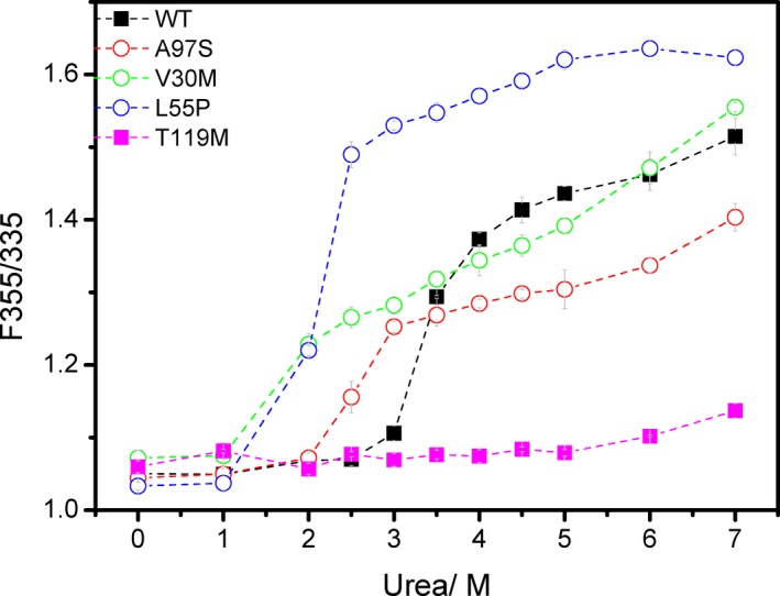
Figure 1.

Urea denaturation monitored by tryptophan fluorescence (F355/F335). WT, wild type TTR, black filled squares; A97S‐TTR, red open circles; V30M TTR, green open circles; L55P TTR, blue open circles; T119M TTR, pink filled squares. Dashed lines are drawn to guide the eye.
