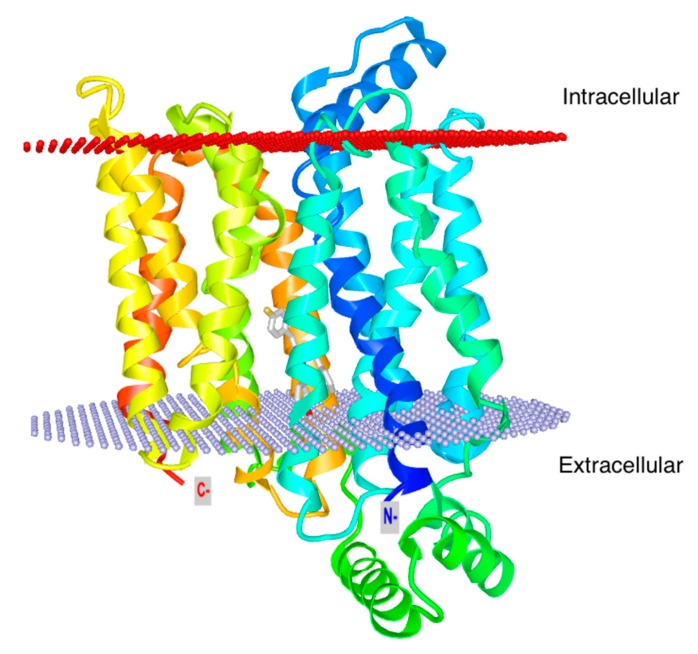
Figure 2.
Glut1 structure. Ribbon model of GLUT1 in the ligand-bound inward facing conformation (PDB: 4PYP; https://www.rcsb.org/structure/4PYP). The N terminus is colored in blue and the C terminus in red. The corresponding transmembrane segments in the four 3-helix repeats are colored the same. The position of glucose bound in the inward facing state is depicted in gray sticks. The structure figure is customized with iCn3D.

