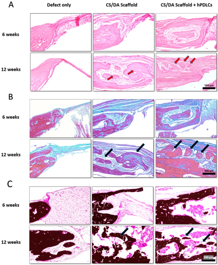
Figure 5.
Effect of CS/DA scaffold on bone regeneration in mouse calvarial defects as assessed by histology (×20). (A) H&E staining shows newly formed bone and dense connective tissue (red arrows) in the group of CS/DA scaffold implanted with hPDLCs and in the group of CS/DA scaffold alone. (B) Masson’s trichrome staining of CS/DA scaffold with and without hPDLCs after 12 weeks showed an increase of blue color representing collagen and mineralized matrix (black arrows). (C) Undecalcified sections with Von Kossa staining of both scaffold alone group and scaffold with hPDLCs group showing intense black mass representing mineralized matrix (black arrows).

