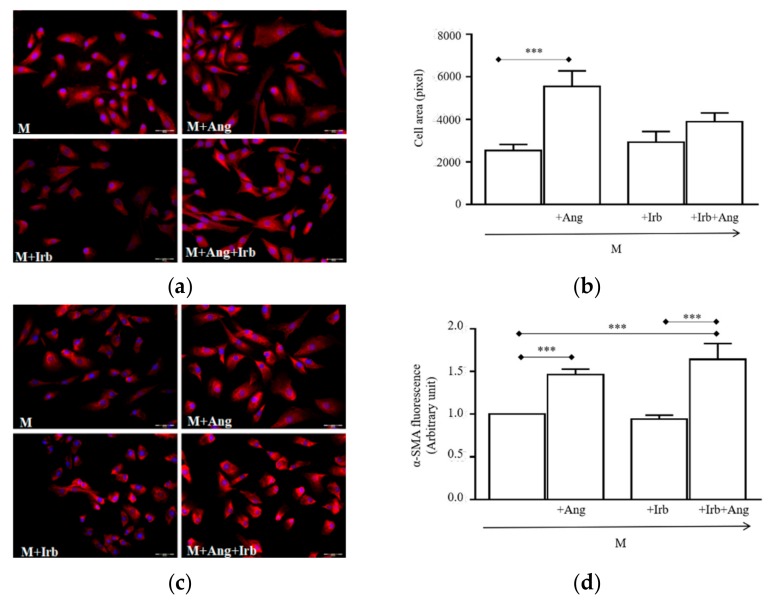
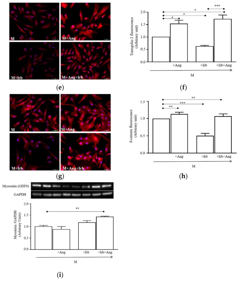
Figure 4.
Effect of sub-chronic conditioning with Ang on hSC-size and myofibroblast markers: (a) Representative photomicrographs of cells labelled with Alexa Fluor (red) and DAPI (blue); (b) Values of cell cross-sectional areas (mean ± SEM) evaluated blind by two researchers using the drawing function of ImageJ software; immunolabelling and relative quantifications of α-smooth muscle actin (c,d), transgelin-2 (e,f), and β-catenin (g,h). Target proteins are stained in red, cell nuclei are in blue (DAPI). Quantifications of protein expression were performed blind by two researchers. All image magnifications are 20×; (i) immunoblotting (top) of myostatin detected in protein extracts of cultured hSCs and densitometric quantification (bottom). * p < 0.05, ** p < 0.01 and *** p < 0.001. In this experiment, hSCs were cultured for 24 h in a standard medium (M) or M supplemented with 100 nM Ang (M + Ang), or 1 µM irbesartan (M + Irb) or 1 µM Irb plus 100 nM Ang (M + Irb + Ang).


