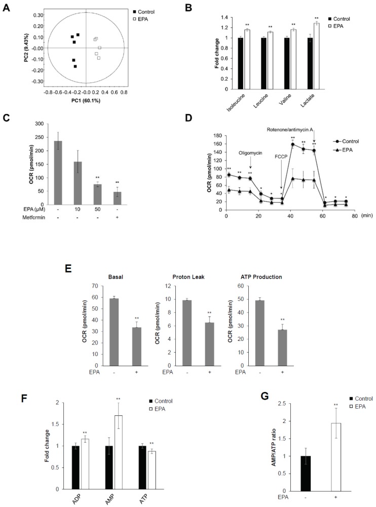
Figure 1.
EPA inhibits mitochondrial oxygen consumption rate (OCR) and decreases intracellular AMP:ATP ratio in skeletal muscle cells. C2C12 cells were stimulated with 50 μM EPA for 3 h. Cellular metabolites were extracted with MeOH/water/CHCl3, and NMR-based metabolic profiling was conducted. (A) PCA score plot for 1H-NMR spectra of cellular metabolite extract levels for metabolome analysis. (B) Levels of BCAAs (isoleucine, leucine, and valine), lactate, fumarate, and malate in C2C12 cellular metabolite extracts. (C) C2C12 cells treated with the indicated dose of EPA and 10 mM metformin for 18 h, respectively. Mitochondrial oxygen consumption rate (OCR) measured using an XF24 analyzer. Metformin was used as a positive control. (D) Mitochondrial OCR in EPA (50 μM) stimulated cells measured using an XFp analyzer in response to 1 μM oligomycin, 0.5 μM FCCP, and 0.5 μM rotenone/antimycin A. (E) Basal respiration, proton leak, ATP production calculated from (D). (F) 1H-NMR-based metabolic profiling analysis showing the intensity of the adenosine phosphates ATP, ADP, and AMP in EPA-treated or untreated cellular metabolite extracts. (G) AMP:ATP ratio derived from 1H-NMR spectral intensities. * p < 0.05, ** p < 0.01 compared to untreated cells. Results from three independently replicated experiments are presented.

