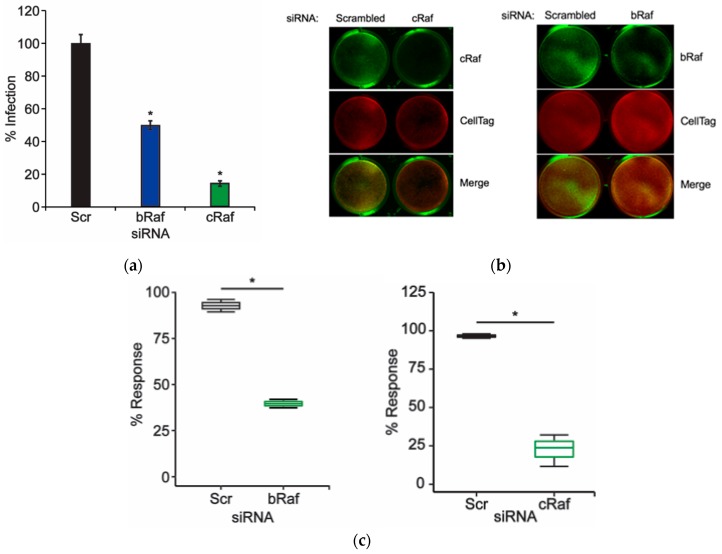
Figure 1.
Knockdown of B-Raf and C-Raf inhibits JCPyV infection. SVG-A cells were transfected with either a scrambled (Scr) siRNA control or a B-Raf or C-Raf siRNA and incubated at 37 °C. At 72 h post-transfection, siRNA-transfected cells were either (a) infected with JCPyV (MOI: 1 FFU/cell) at 37 °C for 1 h and then fed with cMEM and incubated for 72 h or (b,c) processed for ICW analysis of protein knockdown using B-Raf or C-Raf antibodies (green) and CellTag control (red). (a) Infected cells were fixed and stained to analyze nuclear JCPyV VP1 expression. Data are representative of the mean percentage of JCPyV VP1+ cells per 10x visual field normalized to the siRNA control cells (100%) for triplicate samples (representative of three independent experiments). Error bars = SD. (b) B-Raf and C-Raf protein expression was measured for control- and experimental-siRNA treatments by ICW analysis, and (c) signal intensity values were quantified per the calculation [(protein/Cell Tag) × 100] as determined through ImageJ analysis. Boxes represent the distribution of the data for three independent experiments; box midline represents the median. Whiskers represent values 1.5 times the distance between the first and third quartiles (inter-quartile range). Student’s t-test was used to determine the statistical significance. *, p < 0.05.

