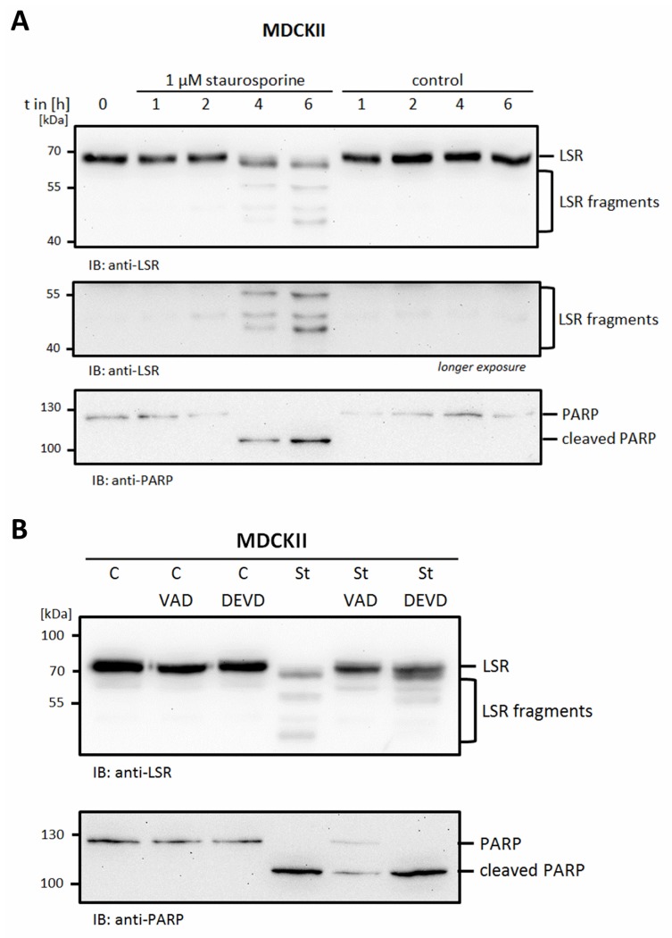
Figure 5.
LSR in apoptotic cells. (A) Treatment of MDCKII cells with 1 µM staurosporine or solvent control for different times as indicated induced fragmentation of LSR. (B) Pre-treatment with caspase inhibitors (10 µM Z-VAD-FMK or 20 µM Z-DEVD-FMK for 1 h) inhibited fragmentation induced by staurosporine (1 µM for 6 h). Cell lysates were analyzed by Western blotting with anti-LSR and anti-PARP antibodies. Representative images of three independent experiments are shown.

