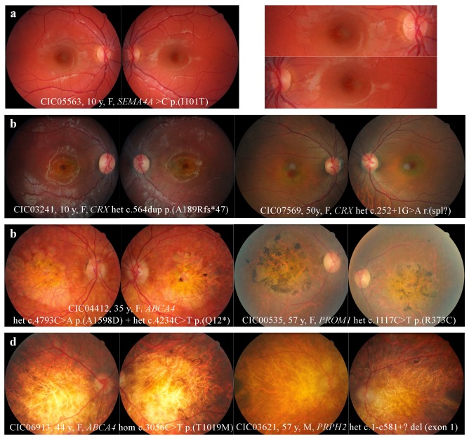
Figure 1.
Fundus abnormalities observed in cone and cone-rod dystrophy (COD/CORD) patients of the cohort. (a) Macular retinal pigment epithelium (RPE) alterations. (b) “Bull’s eye maculopathy” defined by perifoveal atrophy sparing the fovea. Note the temporal pallor of the optic disc for patient CIC03241, a clinical feature known to be associated with CODs. (c) Retinal and RPE atrophy limited to the macular region. Note the pigmented aspect above the macular atrophy, sharply marked in patient CIC00535. (d) Extensive retinal atrophy from the macula to the peripheral retina. Note the optic disk pallor, narrowing vascular network, and peripheral osteoblasts evoking the differential diagnosis of retinitis pigmentosa.

