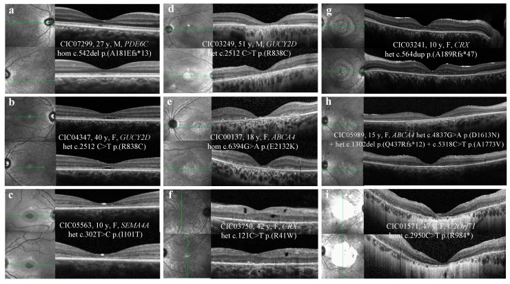
Figure 2.
SD-OCT abnormalities observed in COD/CORDs patients of the cohort. (a) Abnormalities limited to the foveal region, irregular aspect or disruption of the ellipsoid zone (EZ) and the interdigitation zone (IZ). (b) Hyporeflective foveal cavitation. EZ and IZ are disrupted, while ELM and RPE layers are respected. (c) Perifoveal and foveal abnormalities. EZ and IZ are disrupted, while ELM is respected. (d) Outer retinal atrophy of the foveal and perifoveal regions. (e) Hyper-reflective deposits above the RPE in the foveal and perifoveal regions. (f) Hyporeflective cysts at the level of the outer and inner nuclear layers without macular edema. (g) Foveal sparing of the outer hyper-reflective layers; visual acuity is quite preserved for this patient (20/63 OD, 20/80 OG). (h) Outer retinal atrophy of the macular region. (i) Extensive chorioretinal atrophy with retinal thinning of the foveal region and choroidal hyperreflectivity by window defect. SD-OCT: spectral-domain optical coherence tomography; COD/CORDs: cone and cone-rod dystrophy; EZ: ellipsoid zone; IZ: interdigitation zone; ELM: external limiting membrane; RPE: retinal pigment epithelium.

