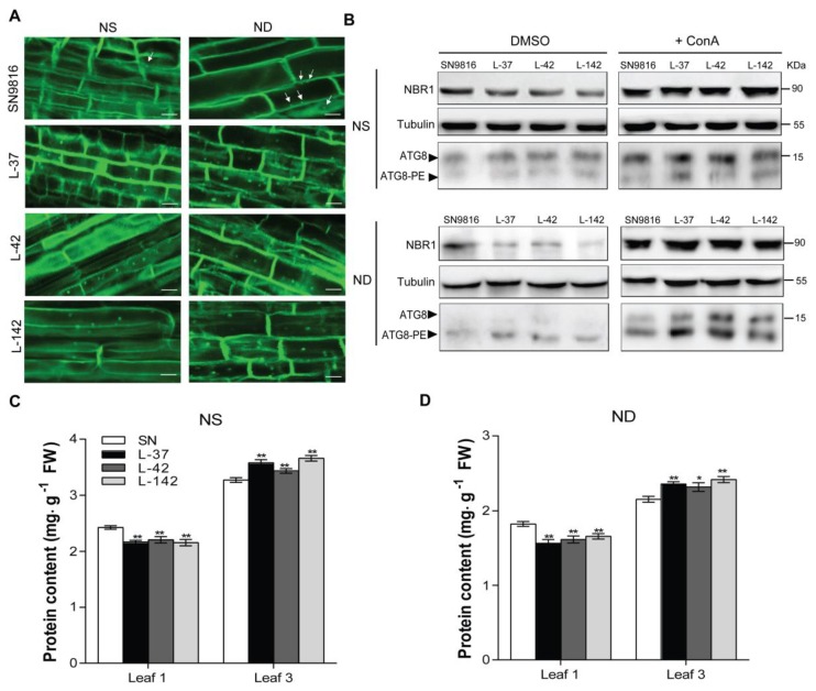
Figure 4.
The overexpression of OsATG8c enhanced the autophagy levels and affected leaf protein profiles in rice. The 21-day-old seedlings of SN9816 and overexpressors were transferred to NS (3.5 mM N) or ND (0 mM N) liquid medium for 24 h. (A) Confocal analysis of autophagosomes in roots by monodansylcadaverine (MDC) staining. MDC-labeled structures are shown in green. The white arrowheads indicate the MDC-stained autophagosomes. Scale bars: 10 μm. (B) Fourteen-day-old seedlings were transferred to NS (3.5 mM N) and ND (0 mM N) liquid media with 0.5 µM concanamycin A (ConA) or solvent control DMSO for 24 h. Immunoblot analysis was used to determine the accumulation of NBR1 with anti-NBR1 in Arabidopsis; near-equal protein loads were confirmed with an α-tubulin antibody. The membrane fraction was used to detect lipidated (ATG8-PE) and free ATG8 levels with anti-Arabidopsis ATG8a in leaves. (C) The protein content of old leaves (Leaf 1) and (D) young leaves (Leaf 3) in SN9816 and transgenic rice under NS or ND conditions. The protein content in equal weight leaf samples was determined by bicinchoninic acid (BCA) protein assay, which was represented as mg protein/g fresh weight. Values are the means ± SD (n = 3), * p < 0.05, ** p < 0.01 (t-test).

