Figure 5.
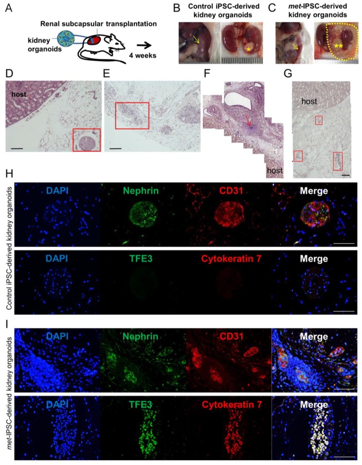
Analysis of tumors generated by transplantation of kidney embryoid bodies. (A) Experimental protocol used. (B,C) Macroscopic aspect of kidney NSG mouse 1 month after transplantation of control (*) or met-IPSC-derived kidney embryoid bodies (**), mouse kidney (arrow). (D,H) Pathological analysis of tumors generated by transplantation of control kidney embryoid bodies at 1 month showing the presence of normal embryoid bodies-like structures expressing kidney differentiation markers. (E–G,I) Pathology of c-met-mutated tumors after transplantation of met-IPSC-derived kidney embryoid bodies at 4 weeks, showing disorganized structures with expression of kidney cancer markers TFE3 and cytokeratin 7. Immature cartilage (arrow). Scale bar: 100 μm.

